A research collaboration between the Cockcroft Institute (including its stake holding partners: University of Liverpool Physics department and STFC Daresbury Laboratory’s ASTeC department), the Royal Liverpool and Broadgreen University Hospitals NHS Trust, the National Research Council ISM of Rome Tor Vergata and the Institute of Translational Medicine have recently collected the first scanning near field optical images of oesophageal cancer using the InfraRed Free Electron Laser (IRFEL) on the ALICE accelerator at Daresbury.
Oesophageal cancer is the fastest rising cancer in the western world. The current understanding of the development of this disease has reached a point where there would be considerable improvements in both diagnosis and prognosis from the information that would be revealed by the application of advanced imaging technologies in the infrared. The aim of the collaboration is to develop a diagnostic test by imaging tissue obtained by endoscopy from patients with a precursor condition called Barrett’s oesophagus
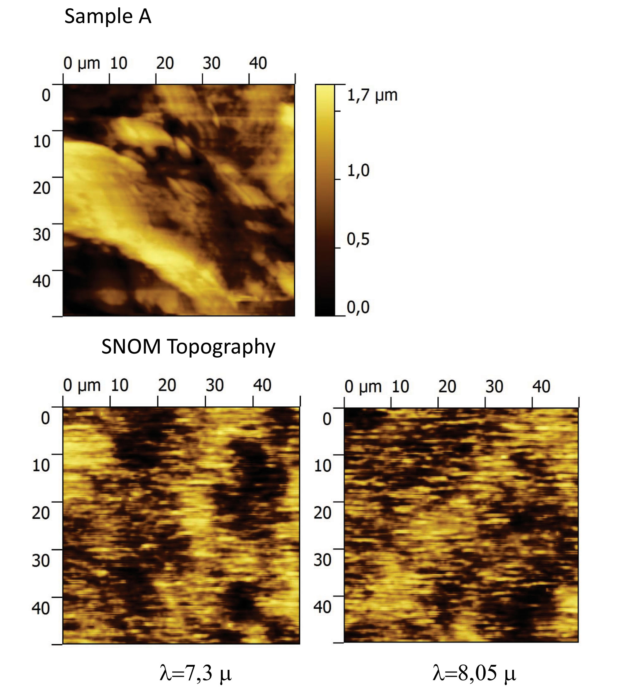
The images above show a topographical image obtained using atomic force microscopy and the same area imaged using 8.05 micron radiation which indicates the distribution of DNA and 7.3 micron radiation which indicates the presence of glycoproteins. The differences in these images demonstrate that although the image is noisy and relates to only a small area of the specimen it will be possible to develop a diagnostic test for the development of oesophageal cancer based on this technique.
The collaboration are now developing improved instrumentation capable of obtaining images from large areas of specimens and with improved signal to noise.